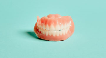
For almost 100 years, any patient requiring teeth replacement, meaning partials, bridges, or complete removable dental prostheses, only had one option—milled dentures. A denture base is a clear, pourable, acrylic resin, made of a product called polymethyl methacrylate (PMMA) that comes in a puck-like form. A fabricator or lab then mills and carves the artificial teeth from the resin.
Manufacturing these prosthetics took a significant amount of time and several visits by the patient to the dental office among other inconveniences that we, at the time, all considered normal. Then came the introduction of computer-aided design, manufacturing, and 3D printing of a denture base and teeth. Since the integration of modern technology into dentistry, there have been improvements in:
- Time in the chair time for the patient
- Time chairside for the dentist
- Number of appointments
- Quicker denture delivery
- Record-keeping
- No-appointment replacements
- Retention of fabricated prosthetics
- Clinical and patient-improved results and satisfaction
With the advent of intraoral scanners, computer-aided design, and the introduction of more modern technology into dentistry, chair time, delivery time, and even precision of prosthetics have been greatly increased. With a digital scan, the measurements of a patient’s mouth and dentures needs can be delivered instantaneously. With 3D printed dentures, the actual prostheses can be produced at a greater degree of accuracy, and then produced equally fast. It’s cheaper and faster, and many patients and clinicians prefer the results. But is it better? Let’s investigate.
Milled denture impression
Milled dentures were originally completely analog. Patients provided a bite impression at their dentists’ office, and then dentures were created based on a plaster mold made from that impression. The process was messy, with the patient enduring a first impression of the existing teeth using alginate or polyethers, polyvinyl siloxanes, and hybrids.
Choosing the right impression material is also a matter of personal preference. Most dentists use alginate, the go-to material in the dental world, due to cost, speed, and ease of use, but some prefer silicone impression material, which they say captures more detail, which is good for crowns, bridges, and restorations.
Alginate distorts easily, so casting must be done quickly, meaning dental offices need to send their impressions and casts to the dental laboratory with expedience—especially compared to the more durable, longer-lasting silicon.
Of course, silicon also has its detractors. While the material picks up more grooves and spaces, it’s very thin, making it far more unpleasant for the patient—especially if it finds the back of their throats. Those with intense gag reflexes need not apply. Silicon also shrinks when curing, so it can’t be used for a full-mouth cast.
Dentists make two rounds of impressions. In the first, material is placed in stock trays that come in generic sizes. Of course, all mouths are different, so stock trays are only the first step. This round is done so that dentists can fabricate a custom-made impression tray for the second round.
These are referred to as “Wash Impressions” or “Border Molding.” During this phase, dentists will use a “Light Body” or “Heavy Body” Wash impression on the customer tray. This Wash is meant to capture patient muscle movement and the anatomy of their mouth.
The second impression is intended to be more precise, because it’s the one sent to the lab to make dentures. But it doesn’t end here: the lab will typically use the Wash impression to fabricate something called Occlusal Wax Rims, and send these back to the dentist’s office for comparison to the patient. As you can see, this is a tedious, complicated process that involves multiple visits from a patient before a lab even begins to fabricate restorations.
And still, impression material capabilities do not always meet every patient’s needs. Some are not precise enough, because it’s hard to always take perfect molds. And when you go through multiple rounds of a process, you introduce more and more opportunities for human error.
For almost fifty years, little changed. Many doctors and technicians are still going through the same process of heating metal spatulas, shaping wax by hand, converting that wax into acrylic, creating a plaster mold, boiling out that plaster mold, and packing the plaster mold again without a guarantee of complete accuracy. It is time-consuming and requires (expensive) manual labor.
Patients want less time in the chair, and so does the dentist. Patients also want restorations and replacement teeth to fit. Imagine multiple, unending dentist visits for uncomfortable impressions, followed by receiving a prosthesis that makes your mouth and gums irritated or inflamed, or even injures existing jawbone — all because of fifty-plus year old manual technology. We haven’t even added teeth to these fitting, but you can already see why many dentists (and us at Dandy) are excited about the future of intraoral digital scanning.
Adding teeth to milled dentures
We have described the back-and-forth with the lab on the plaster molds and impressions, but we’ve yet to add teeth to these fittings.
The synthetic resin we’re using (a solid block disc of polymethyl methacrylate, aka PMMA) is durable and strong. It is found in denture bases and artificial teeth, but also in shatterproof windows, skylights, and aircraft canopies. Considering that the average human bite strength is 162 pounds per square inch (psi), with the second molars exerting a bite force between 1,100 and 1,300 Newtons, you can understand why patients may lose 70% of their bite force with traditional dentures, and why many dentists opt for something as strong as PMMA. With replacement teeth that are this strong, anatomy and fit matters.
So once the wax impression has been returned to the lab, it is prepared for the final process. Each manufacturer has its own methodology, but the standard process as prescribed by the Foundation for Oral-Facial Rehabilitation is that the dentures are removed from the articulator mountings and inspected for any damage. They are then placed in water and then positioned in a flask, which is then partially filled with stone. The positioning within the flask must land the cast and plaster at the same level as the edge of the flask. Then the wax is replaced with polymerized acrylic resin.
Okay, now let’s add the teeth.
Modern milled dentures typically have two types of artificial teeth: carded or milled. They are chosen to be compatible with the patient’s structure of face, parfunction, previous dentures or partials, and jaw relationship. Carded teeth, named for the identification card they were originally placed on, are identified by mold, shape, and shade of the teeth (and described as versions A, D, C, E, and F). Most carded teeth consist of an anterior or posterior tooth selection guide with both upper and lower teeth.
These teeth are manufactured in an extrusion molding process in a continuous strip. Manufacturers can also include a kit with a “facial meter,” which measures facial features for distance to correlate cards with a variety of different-sized upper teeth. This helps the dentist and patient evaluate and select teeth based on look and feel with the entire face.
Some of the most commonly used are: Ivoclar DCL teeth, a highly durable nanohybrid composite version of acrylic resin, and Dentsply Portrait interpenetrating polymer network (IPN) teeth. Both are premium teeth known for long-lasting wear, and resistance to cracks and chips. And depending on budget and bite strength, versions of artificial teeth in a more economical porcelain are available.
When choosing between either acrylic or porcelain teeth for milled dentures, it comes down to durability, esthetics, and costs. And despite all these considerations, we’re still providing patients a replacement method with some risks. There can always be bonding issues if there are errors in the impression or manufacturing, and given that each tooth or group of teeth is fitted individually, there is always a greater chance of tooth loss than with something printed. Without proper fit or consistent wear of the complete denture, there can be bone loss, as the gums and jawbone end up not being stimulated to provide continued growth, and improper oral hygiene can lead to the deterioration of the dentures and staining of the prosthetic.
Enter tech-aided 3D printed dentures
In the 1980s that computer aided design and manufacturing (CAD/CAM) finally made its way to dentistry. Initially seen as a fad, it has now become a more accepted and useful part of many dentists’ workflow and dental lab work.
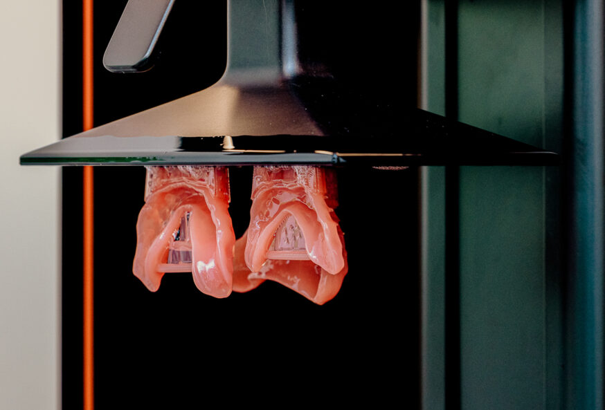
Denture materials initially brought CAD/CAM in, like the Ivoclar Ivobase CAD milling system’s addition of fibered “veins” in the base for aesthetic enhancement. These denture materials looked great, but cost a lot more. When 3D printing found its way to the milling machine, dentistry moved more fully into the digital age.
Still, at the onset 3D printed dentures were honestly not superior. 3D construction had not reached the quality present in milled dentures, because manual care can be precise (and because the method had a 50+ year head start).
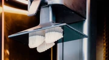
But the technology has caught up, and the second and third generations of teeth have improved massively, as the process has made CAD-milled bases and 3D printed dentures more commonplace. This digital milling saves time and expense, as rapid prototyping can remove weeks from the process of mailing and shipping impression bases back and forth to labs. Studies are also finding that intraoral scans of “periodontally compromised teeth bring a significant advantage for the treatment planning and delivery of outstanding complete dentures.”
CAD/CAM is also passing comparison studies (like this one on PubMed) with flying colors, dismissing and demystifying claims of milled dentures maintaining superiority. When it comes to precisely fabricating teeth replacements, the machines are winning and 3D-printed dentures are becoming a new standard.
Denture printing and digital dentistry
Remember that once a tooth model has been made, the milled-denture patient still has a lengthy journey ahead. The first impression makes a cast, then a second impression, and more visits—usually a minimum of five in-chair visits with possible follow-up consultations for adjustments.
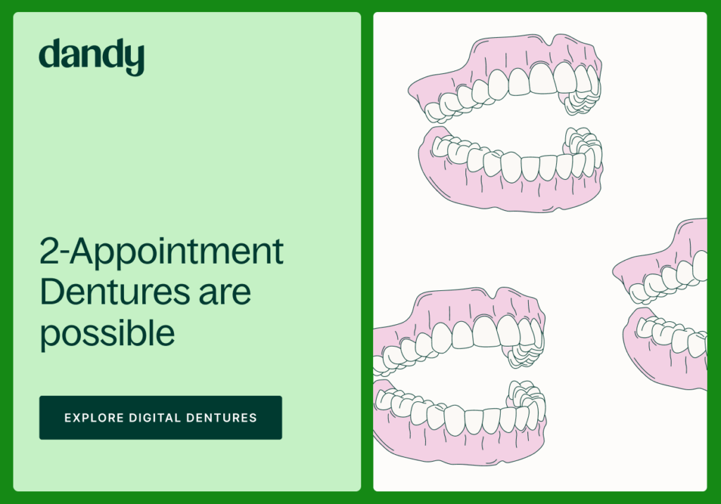
With 3D printed dentures and advances in intraoral scanning, digital dentistry is winning the hearts, minds, mouths, and checkbooks of tooth-replacement patients. With an adept practitioner, and a quality scanner, a patient can be done in 2 visits. That’s 40% of the standard visit, for those keeping score. Add “no gagging” to the scorecard too, as the only thing to clean after visits is the scanner. Gone are the days of goo and hot spatulas.
As technology advances with each iteration of scanners and software, the detail of the scans produces better images, and each can be saved and easily reproduced. (Not to mention: files can be stored on hard drives and the cloud instead of endless shelving or storage spaces).
And we’re also at the advent of exploring new materials with 3D printed dentures and replacements. A study examining the dimensional accuracy and the surface topography of a custom-designed, 3D-printed zirconia dental implant found remarkable accuracy. These kinds of advancements in precision fabrication, combined with even more durable material, threaten to eliminate old headaches like the shrinkage of partials and removable complete dentures.
And as for partial dentures, it is a whole different game, but progress may get the industry to 3D-printed partials soon.
As we’ve outlined, however—it is not the printing of the digital dentures that is the miracle. Fine milled replacements have found their homes in happy smiles for more than a century, and milling works. The digital denture process, including digital scanning and CAD/CAM technology is the real advancement, eliminating time, money, postage, operator error, storage, and patient discomfort. That 3D printed dentures are as good or better than their milled counterparts is only part of an equation that includes constant improvements to partials, dentures, and implants relatively soon, but also to the industry at large — improving the trueness, structure, and flexural properties of dental prosthetics is only one (good) outcome.
There is better accuracy in design, meaning less time in the chair. This also leads to fewer repeat visits and refittings for dissatisfied patients. This brand-new patient experience leads to word-of-mouth marketing, and gives dentists competitive advantage. Scanning (and not dealing with molds, trays, and mail) improves dental office digital workflows and cuts down on chair time by about 15 to 20%. This allows dentists to increase their volume of patients without sacrificing quality of care. When you look at the practice, the patients, the employees, and process, the only question worth asking is not “is 3D printed better,” but instead “what about 3D printed dentures isn’t better?”